Vision-Language Models in Medical Imaging: How VLM Architectures Are Outperforming Radiologists in Early Cancer Detection
The question is no longer whether AI can detect cancer. The question is where and by how much it outperforms human radiologists. Vision-language models (VLMs) are a class of multimodal AI that combin...
What Vision-Language Models Mean for Cancer Detection Today
The question is no longer whether AI can detect cancer. The question is where and by how much it outperforms human radiologists.
Vision-language models (VLMs) are a class of multimodal AI that combine visual understanding with language-based reasoning. Unlike older computer vision models that simply classify images, VLMs process imaging data alongside clinical context — patient history, lab results, prior scans — and generate natural-language descriptions of findings.
By late 2025, over 1,000 FDA-approved AI imaging tools were in active clinical use. The global market for AI in medical imaging reached $2.01 trillion and is projected to hit $22.97 trillion by 2035. Behind the numbers is a practical shift: VLMs are moving from research labs into hospital infrastructure, and the performance data is becoming hard to ignore.
This article breaks down what VLMs actually do, how they compare to radiologists across cancer types, where the technology falls short, and how health systems are deploying it today.
The VLM Architecture Advantage Over Single-Modality AI
Traditional computer vision models for medical imaging rely purely on pixel-level pattern recognition. A CNN (convolutional neural network) trained on chest X-rays learns to spot lung nodules. It has no way to incorporate the patient's smoking history, the radiologist's prior read, or the clinical note saying "rule out malignancy."
VLMs solve this with a dual-encoder design:
Vision encoder — processes the imaging data using a vision transformer (ViT) or CNN backbone. Converts X-rays, CT scans, MRI, and pathology images into feature vectors.
Language model — takes those visual features and reasons over them using a large language model. This component is what enables clinical contextual reasoning and natural-language output.
Multimodal fusion — combines visual and textual representations, allowing the model to correlate imaging findings with clinical context. A VLM analyzing a mammogram can weight findings against the patient's age, family history, and prior imaging.
This architecture is why VLMs consistently outperform single-modality models on complex diagnostic tasks. The language model provides reasoning capacity that pure visual classifiers lack.
VLM Performance vs. Radiologists: The Evidence
Here is what peer-reviewed and real-world studies show across the most common cancer detection use cases.
94.5% accuracy — Google's AI mammography system achieved this across a large clinical validation dataset. False positives dropped 5.7%. False negatives dropped 9.4% compared to radiologist-only reads.
The German real-world study was more striking. Across 463,000 women screened with AI-assisted mammography, breast cancer detection increased 17.6% compared to standard-of-care radiologist reading. No lab is more compelling than a population-scale real-world result.
Lung cancer screening with low-dose CT (LDCT) shows similar patterns. AI nodule detection systems consistently hit 85-94% sensitivity across large screening cohorts. On high-volume screening workloads — where radiologists read hundreds of scans per shift — AI maintains consistent performance. Human radiologist accuracy measurably declines during extended sessions, a phenomenon known as "decision fatigue."
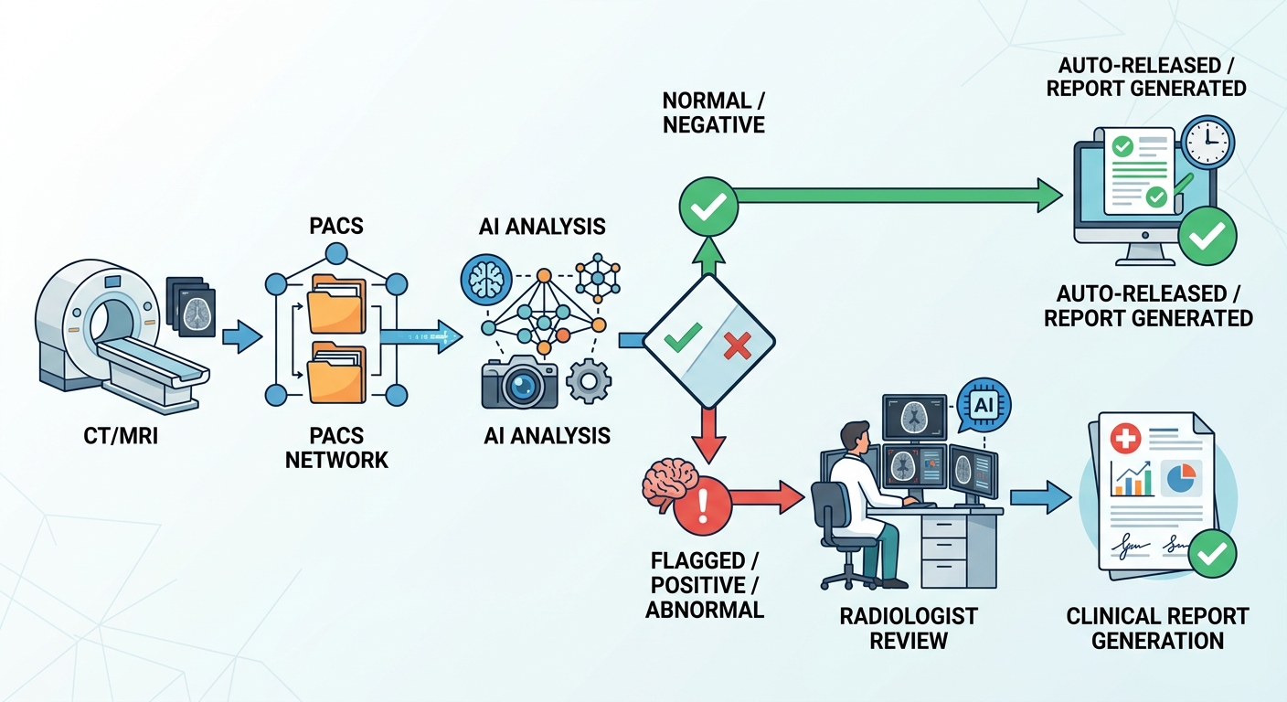
The "second reader" model is where the data gets most interesting. When AI acts as a first-pass pre-screener — auto-releasing normal scans and flagging suspicious ones — radiologists can focus their expertise on cases that actually need it. Studies show this hybrid approach:
- Increases cancer detection rates 8-12%
- Reduces radiologist workload approximately 25%
These are not hypothetical projections. They are documented outcomes from programs running in clinical settings today.
Where AI Gets It Right and Where It Still Struggles
VLMs are not infallible. Understanding both sides of the ledger is critical for responsible deployment.
Where VLMs reliably outperform humans:
- Processing high volumes of routine screening cases consistently
- Detecting subtle patterns in early-stage lesions that human eyes may miss under fatigue
- Cross-referencing imaging findings against large-scale population data
- Maintaining stable performance across extended shifts and high caseloads
Where VLMs still fall short:
- Rare cancer subtypes with limited training data
- Atypical presentations that diverge from common patterns
- Cases where clinical context is ambiguous or conflicting
- Hallucination — generating plausible but incorrect findings in text output
- Algorithmic bias — underperformance on demographic groups underrepresented in training data
Fully autonomous clinical diagnosis is not the current standard. VLMs function best as decision-support tools that augment radiologist expertise, not replace it.
The Radiologist Shortage: Why AI Assistance Is a Workforce Solution
The performance numbers matter for another reason: there are not enough radiologists.
Demand for diagnostic imaging is growing faster than the radiologist workforce globally. Screening programs for breast, lung, and colorectal cancer are expanding. Aging populations increase imaging volume. Meanwhile, radiology training pipelines are constrained.
AI addresses both sides of this mismatch. By automating the "easy" reads — normal or clearly benign cases — radiologists can redirect their expertise to complex diagnostics, equivocal findings, and cases requiring nuanced clinical judgment.
The 25% workload reduction documented in AI-assisted screening programs is not about replacing radiologists. It is about making the existing workforce more effective. A radiologist reviewing 60 flagged cases instead of 200 routine scans brings more attention and expertise to each one.
For health systems, this translates directly: better detection rates, faster turnaround for patients, and more efficient use of high-cost specialist time.
Key Cancer Types Where VLMs Show Strongest Results
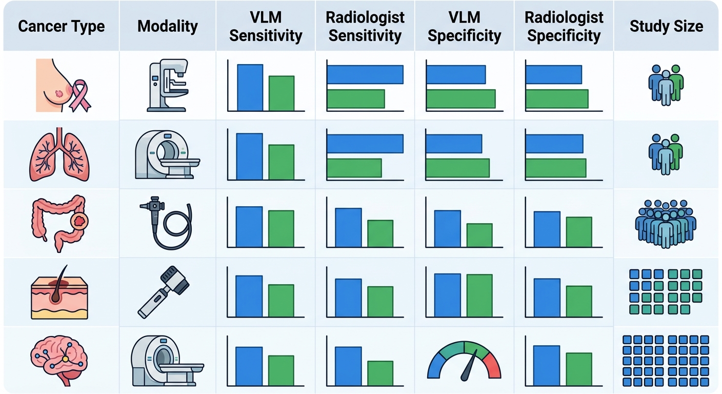
Breast Cancer
The strongest evidence base for VLM deployment. Google, Lunit, and iCAD all have FDA-cleared or CE-marked AI mammography tools. The 463,000-woman German study remains the landmark real-world result: 17.6% detection improvement with AI as second reader.
Lung Cancer
Low-dose CT lung cancer screening has a well-established AI workflow. Nodule detection sensitivity of 85-94% on LDCT is documented across multiple vendor platforms. The National Lung Screening Trial (NLST) data provided the evidence base; AI tooling has since built on top of it with improved specificity.
Colorectal Cancer
AI-assisted colonoscopy with real-time polyp detection is one of the fastest-growing deployment areas. VLMs trained on colonoscopy video frames flag suspicious mucosal lesions in real time. Adenoma detection rate improvements of 10-15% have been reported in meta-analyses.
Skin Cancer
Dermoscopy AI uses VLM architectures trained on large image databases (ISIC archive) to classify pigmented lesions. Performance is approaching dermatologist-level sensitivity for melanoma detection in controlled settings.
Brain Cancer
MRI-based VLM systems are being trained to identify suspicious masses and predict progression patterns. This area has more limited real-world deployment but active research investment.
Deploying VLMs in Hospital Infrastructure: Practical Considerations
Health systems evaluating VLM deployment need to understand the technical and regulatory landscape. Here is what the implementation process actually looks like.
Integration with existing systems — Most hospital imaging infrastructure runs on PACS (Picture Archiving and Communication Systems) and RIS (Radiology Information Systems). VLM platforms connect via DICOM (for imaging data) and HL7/FHIR (for clinical context). Most enterprise platforms provide REST API endpoints for integration, and most PACS vendors support standard DICOM interfaces.
On-premise vs. cloud — Protected health information (PHI) creates data residency constraints. Many health systems prefer on-premise or hybrid deployments where imaging data never leaves their infrastructure. Cloud deployment is viable when data is de-identified or when the cloud provider offers HIPAA-compliant infrastructure with BAA agreements.
Validation requirements — Before clinical deployment, VLMs must undergo internal validation (on institutional data) and external validation (on data from other populations). This is where most academic models fail: trained on one hospital system's data, they underperform when tested on another. Rigorous external validation on diverse populations is the minimum standard.
FDA clearance — Most VLM medical imaging tools fall under FDA 510(k) clearance as Class II medical devices. As of 2025, over 1,000 AI imaging tools had received FDA clearance. The regulatory pathway is well-established but requires documented clinical validation studies.
Production monitoring — Once deployed, VLMs need drift detection and performance tracking. Model accuracy can degrade as the patient population shifts, imaging protocols change, or hardware is updated. Continuous monitoring against ground truth outcomes is essential.
Fine-Tuning VLMs on Medical Imaging Data
General-purpose VLMs can be fine-tuned on institutional data for specific modalities or cancer types. The process involves:
- Starting with a foundation model (Med-PaLM, BioMedCLIP, RadFM, or similar)
- Fine-tuning on the health system's own labeled imaging data
- Evaluating on held-out institutional data to check for overfitting
- External validation on a different patient population
Common pitfalls include dataset leakage (training data appearing in validation set), demographic bias in training cohorts, and overfitting to institutional imaging protocols that do not generalize.
Leading VLM Platforms for Medical Imaging in 2026
The market is actively consolidating around several categories of players:
Foundation model providers — Google Med-PaLM M3, Microsoft Azure AI Health, and Anthropic/clinical partners are building multimodal models with strong medical visual question answering capabilities.
Enterprise imaging AI platforms — Aidoc, Zebra Medical Vision (now Nanox AI), Qure.ai, and Lunit offer FDA-cleared tools across multiple imaging modalities with PACS integration.
Open-source models — BioMedCLIP, RadFM, and CheXNet-derivative models provide research-grade VLMs for institutions that want to build and fine-tune their own models.
Comparison criteria — When evaluating platforms, health systems should prioritize: documented clinical accuracy on their target modality, FDA clearance status, integration complexity with existing PACS/RIS, and total cost of ownership including infrastructure and maintenance.
The right choice depends heavily on the use case, existing infrastructure, and institutional data strategy.
The Future: Human-AI Collaboration in Radiology by 2030
The trajectory is clear. Within the next five years:
AI will handle first-pass screening for routine cases across most high-volume imaging modalities — mammography, chest X-ray, CT nodule screening. Radiologists will shift to complex diagnostics, equivocal findings, and cases where clinical context is critical.
VLMs will become more explainable. Current models generate natural-language findings, but the reasoning process behind those findings is largely opaque. The next generation of models will produce structured justifications linked to visual evidence.
Regulatory frameworks will adapt. As AI becomes standard-of-care in radiology, reimbursement frameworks will evolve to reflect AI-assisted diagnosis and the productivity gains it delivers.
What will remain human: clinical judgment on complex presentations, patient communication, rare disease expertise, and accountability for final diagnoses. The radiologist of 2030 will be a more strategic interpreter of imaging data, not a bulk reader of routine scans.
Expert Q&A
Q: Can VLMs fully replace radiologists in cancer detection? A: No. Current VLMs work best as decision-support tools — a second reader that flags findings and prioritizes cases. They excel at high-volume routine screening but require human oversight for complex diagnoses, unusual presentations, and cases with ambiguous clinical context. Fully autonomous clinical diagnosis is not the standard of care.
Q: What is the main advantage of VLMs over traditional computer vision models? A: Multimodal reasoning. VLMs combine visual pattern recognition with language-based analysis, incorporating clinical context — patient history, lab results, prior imaging — into diagnostic output. This produces more accurate and contextually appropriate findings than purely visual classifiers.
Q: How accurate are VLMs for early cancer detection compared to experienced radiologists? A: VLMs achieve 85-94% sensitivity for lung nodule detection, 94.5% accuracy for breast cancer on mammography, and consistently match or exceed radiologist performance at scale. Critically, AI maintains consistent accuracy across thousands of scans, while human accuracy degrades during extended reading sessions.
Q: What are the biggest risks of relying on VLMs for cancer detection? A: Hallucination (AI generating plausible but incorrect findings), algorithmic bias (underperformance on underrepresented demographics), and over-trust leading to missed edge cases. Robust validation protocols, diverse training data, and mandatory human oversight are essential before clinical deployment.
Q: How do hospitals integrate VLM cancer detection into existing workflows? A: Integration happens via API connections to PACS/RIS using DICOM and HL7/FHIR standards. Most enterprise platforms support cloud-hybrid deployment with on-premise inference for PHI-sensitive imaging. The process involves validation, FDA clearance review, integration testing, and production monitoring.
Q: What is the difference between VLMs and standard CNN-based AI in medical imaging? A: CNN-based AI (e.g., CheXNet for chest X-rays) classifies images using pixel-level pattern recognition only. It outputs a probability score for a condition. VLMs add a language model component that reasons over visual features in context, enabling natural-language explanation of findings and integration with clinical text. A VLM can answer "what are the three most suspicious findings in this mammogram and how do they compare to prior imaging?" — a capability CNNs do not have.
Q: How does VLM performance vary across different imaging modalities — CT, MRI, pathology? A: CT imaging has the most mature VLM evidence base, particularly for lung nodule detection and mammography. MRI VLMs are advancing quickly but have less real-world deployment data. Digital pathology (WSI — whole slide imaging) is an emerging frontier where VLMs show promise for cancer grading and biomarker identification, but slide-sized images create significant computational challenges. The modality with the most FDA-cleared tools as of 2026 is mammography, followed by chest CT and X-ray.
Q: What does "hallucination" mean in the context of VLM medical imaging, and how is it mitigated? A: Hallucination in VLMs refers to the model generating plausible-sounding but factually incorrect text output — for example, describing a finding that is not present in the image, or mischaracterizing a lesion's properties. Mitigation strategies include: output constrained decoding (limiting the vocabulary the model can output), retrieval-augmented generation (grounding outputs in verified references), confidence thresholding (suppressing low-confidence outputs), and mandatory human review of all AI-generated text. No current clinical deployment should rely on unreviewed VLM text output.
Q: How do you evaluate whether a VLM will generalize from one hospital to another? A: Generalization testing is the critical gap in most VLM publications. Before deployment, a VLM should be tested on: (1) held-out institutional data from the same hospital (internal validation), (2) data from different hospitals and geographic regions (external validation), (3) data from different scanner manufacturers and imaging protocols, and (4) data with different demographic distributions. Performance drops in external validation are common — a model that hits 94% accuracy in a Google internal study may drop to 87% on a different hospital's data. Any vendor claiming "trained on millions of images" should provide external validation results on populations similar to yours.
Q: Is there a risk that widespread AI use in radiology causes skills atrophy in human radiologists? A: This is a legitimate concern. If radiologists progressively delegate routine reads to AI, their ability to independently read complex cases may degrade over time. Best-practice programs address this by using AI as a "second reader" rather than a "first reader" — the radiologist always reads the case but has AI input. Some programs rotate radiologists off AI-assisted workflows periodically to maintain independent reading skills. The goal is augmentation, not dependency.
Q: What are the most promising emerging directions for VLM medical imaging beyond cancer detection? A: Three directions stand out in 2026: (1) Treatment response prediction — VLMs analyzing serial imaging to predict how tumors respond to therapy; (2) Prognostic modeling — VLMs predicting patient outcomes by integrating imaging with clinical data over time; (3) Multi-cancer screening — unified VLM systems that simultaneously screen across multiple cancer types from a single imaging study. Foundation model research from Google Health, Microsoft Research, and academic medical centers is most active in these directions.
Ready to evaluate AI imaging tools for your health system? Subscribe for practitioner-focused analysis of medical AI deployment, benchmarking, and implementation guidance delivered to your inbox.
Image URLs
| # | Alt | URL |
|---|---|---|
| 1 | VLM accuracy vs radiologist performance comparison across 5 ... | /api/images/b3c620a3dc0841089b6adfb1db92f029 |
| 2 | Radiology AI workflow flowchart showing DICOM image acquisit... | /api/images/aa04292f7ee04998aef426cc73a0de61 |
Total: 2 images uploaded